2024 Update: Brain impacts from the COVID vaccines
mRNA COVID vaccines have been shown to have damaging impacts on the brain. Here’s how that happens. A summary of recent research.
Cognitive damage and Alzheimer’s disease
Male neonatal rats have shown substantially decreased neuron counts and autistic behaviors after COVID vaccine dosing of their mothers. That next generation following vaccination of the mothers (not the neonates) showed, after birth, these traits, as well as impaired motor performance, with reduced coordination. Very sadly, for corresponding human victims, the neuron count in the neonatal male rats was reduced by over 31% compared to controls. The cerebellar Purkinje fiber count, which would be associated with spatial orientation and coordination of body movements, was reduced by 62%. [1] These decreases are not only enormously significant, but are also horrifying when we think of the pregnant women all over the world who were urged, cajoled, even bullied to take COVID vaccines during pregnancy.
Korean researchers Roh, Jung et al gathered public medical records of half of all of the residents of Seoul, South Korea, as of January 1, 2021. The 50% were chosen at random from the entire population of that city. Then from that population, the Roh team examined medical records of those aged 65 years and over. From the medical records of that half of Seoul’s senior population, vaccination status and ICD-10 codes were obtained. This resulted in a senior population of 558,017 individuals, comprised of 519,330 vaccinated individuals and 38,687 unvaccinated. [2] Vaccinated individuals had received either Pfizer, Moderna, AstraZeneca or Johnson & Johnson vaccines, or a combination of any of those.
The diseases of interest were Alzheimer’s disease, dementia in Alzheimer’s disease, mild cognitive disorder, Parkinson disease and vascular dementia. Those individuals with any of those diagnoses during the prior year were excluded from the study.
The figure below summarizes their methods and findings.
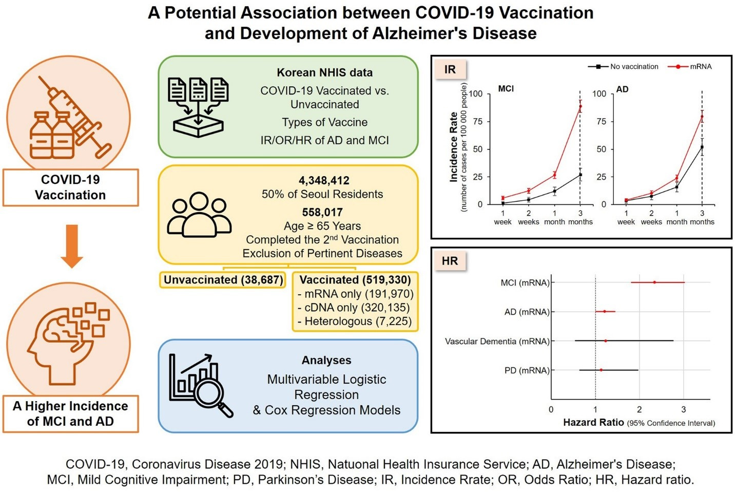
The two graphs on the right of the above figure show more than double the mild cognitive impairment and a higher incidence of Alzheimer’s disease, as discussed below, in the vaccinated population.
The mean age of the unvaccinated was 75.23 and the mean age of the vaccinated was 72.73, yet despite this 2.5 year difference, the vaccinated fared worse in three of the five diseases considered. No significant difference was found for either Parkinson disease or vascular dementia.
For mRNA vaccinated individuals, the hazard ratio for Alzheimer’s disease was 1.209. (95% CI: 1.013-1.444. P = 0.036). So there was a 21% increased chance of Alzheimer’s for the mRNA-vaccinated.
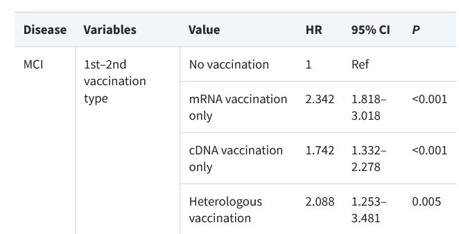
For mRNA vaccinated individuals, the hazard ratio for mild cognitive impairment was 2.342 (95% CI: 1.818-3.018. P < 0.001). This showed that mRNA-vaccinated individuals have more than double the risk of mild cognitive impairment as unvaccinated individuals, even though the average unvaccinated person was older.
The exact hazard ratios (HR, or the odds of acquiring a disease post-vaccine) for mild cognitive impairment are shown below. “Heterologous vaccination” refers to two doses of one mRNA and one cDNA.
Dr. Hiroto Komano is a professor of neuroscience and a researcher at Stanford University. His field of study is molecular analysis of Alzheimer’s dementia. [3] Dr. Komano, commenting on the above study, has expressed his concern that dementia cases will likely increase significantly, especially in the elderly, if there is persistent use of mRNA vaccines. He warns that the current incidence of one in five people over the age of 65 having dementia could double to two out of five, if mRNA vaccination continues. [4]
First, a brief digression to the topic of depression
Significant overlap has been found between depressed and demented patients. After adjusting for demographic and health factors, in the first year after diagnosis, dementia patients had triple the risk of depression compared to those without dementia. [5]
Cognitive and psychiatric effects of COVID vaccination were seen in all ages of adults in the Kim study, which examined the same Seoul Korea population as in the Roh, Jung, et al study. In this study, the Kim team of researchers examined residents over the age of 20, and considered a broader range of mental adverse events. [6] Whether mRNA or DNA vaccines or a combination, the vaccinated cohort showed increased incidence of depression, anxiety and other disorders, compared to the unvaccinated cohort in this population-based study.
At three months post vaccination, the vaccinated cohort showed 68% increased incidence of depression and a 44% increased incidence of anxiety, dissociative, stress-related and somatoform disorders.
The results of that enormous study of two million people were consistent with an earlier and much smaller study by Balasubramanian, [7] in which onset of psychiatric disorders occurred within zero to ten days following the first COVID vaccine dose. Young and middle-aged adults were more commonly affected.
In one literature review, across all adult age groups, mean age 34 years, new onset psychosis was seen to peak within seven days after dosing of the Pfizer vaccines and the ChAdOx1-nC19 vaccines, of which the Astra Zeneca vaccine was one. [8] For one third of the cases, new onset psychosis manifested within one day of receiving the vaccine. This describes a Gompertz curve of temporal effect – skyrocketing incidence at the beginning, followed by gradual attenuation of effect - which is consistent with the temporal criterion of the Bradford Hill criteria for establishing causality.
Post-vaccine inflammation as a cause of both depression and dementia
Here’s why I digressed to discussing depression. It seems that inflammation of the brain is a significant component of both conditions, and there is hardly a substance that has entered the human brain that is more tenaciously inflammatory than the spike proteins. The S1 fraction of the spike protein, spike’s most troublesome part, has been shown to enter the brain. [9] Now when those spike proteins come by way of a mere coronavirus halo (“corona”), as in naturally acquired infection, there are ways to rebuff entry to the brain, such as ivermectin, zinc and hydroxychloroquine’s mechanisms of thwarting ACE2 docking and entry to cells. [10] However, in the case of spike proteins delivered with the state-of-the-art lipid envelopes delivered by the Pfizer and Moderna vaccines, now you have an efficient way to enter the brain that bypasses some of our best strategies against that assault.
The Kim study addressed why depression can be a common finding among the vaccinated. First, correlation between depression and inflammation has been observed for many years. In a different study of over 10,000 people, divided nearly in half between depression patients and controls, such inflammatory markers as Tumor necrosis factor-alpha (TNF-α), interleukins (IL) 3, 6, 12 and 18, as well as C-reactive protein (C-RP) were found to be significantly higher in the patients than in controls. [11] Not only was the difference statistically significant, but the inflammatory markers C-RP and IL-12 were remarkably consistent in the depressed cohort, which further confirmed depression to be a pro-inflammatory state, or to at least have a pro-inflammatory component. There are some common pathways of injury between cognitive injury and depression, and it is worth exploring what contribution, if any, the COVID vaccines contribute to those pathologies.
It should not come as a surprise that increased incidence of both depression and dementia have been observed in COVID-vaccinated patients. Depression and dementia have sometimes been confused in diagnosis, because they both carry many common symptoms, such as apathy and self-neglect, irritability and withdrawal from other people, difficulty with, or lack of effort in, memory and focus. The latter may appear to the clinician as a mental slowness or a cognitive impairment. [12] Sometimes the depressed patient with the above symptoms is said to have a pseudodementia.
Inflammatory effects of spike proteins in the brain
We have seen from cardiovascular effects that there is much of an inflammatory nature about spike proteins and the COVID vaccines. Whether naturally acquired from COVID infection, or by injection from the mRNA vaccines, a similar diffusion of spike proteins are seen throughout the plasma and tissues far and widely dispersed from the injection site, [13] [14] including in the brain. [15] The Buzhdygan team studied the effects of the SARS-CoV-2 spike protein on endothelial cells in the brain, and noted a correlated pro-inflammatory response with effects on the blood brain barrier. [16] At the surfaces of the endothelial cells in the brain that make up the blood-brain barrier, there are pathological changes that arise. [17] [18] This happens by way of degrading of several kinds of proteins that compose the blood brain barrier, and this is provoked by S1 of the spike protein. [19] Those proteins include adherens, tight and gap junctions. And in fact, spike proteins and related debris gather and accumulate there at the endothelial cells that these junctions surround, up to twenty times the accumulation at other cells. [20] This is even before the spike proteins pass that barrier and enter the brain. Once spike proteins are in the brain, they are correlated with neuroinflammatory, pathological changes affecting cognition and mood. [21]
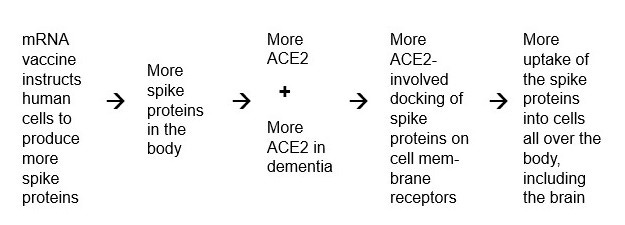
Since 2021, I have discussed cardiovascular effects of spike proteins. https://colleenhuber.substack.com/p/is-it-possible-to-avoid-heart-damage The enzyme ACE2 is increased on cell surfaces in the presence of SARS-CoV-2, and that enzyme serves as a docking station and direct port of entry for the virus to enter a human cell. [22] [23] Hoffman et al describes that process in detail, how the protruding S glycoprotein of the spike protein has a receptor binding domain at which the ACE2 protein docks. [24] Not only is ACE2 present in the cerebral vasculature, at the vascular endothelial cells, as has been known since 2004, [25] but there is also higher expression of ACE2 enzyme in brain tissue derived from demented patients than from controls. [26] In fact, it was at mice brain blood vessels that it was first shown that the endothelial damage seen in SARS-CoV-2, the worst pathology associated with SARS-CoV-2, was caused by the spike protein alone. [27] Furthermore, ACE2 is clearly present in the fronto-cortical regions of the brain, in both capillaries and larger blood vessels. The same study found that dementia patients had an average of 3.5 times as much ACE2 expression as normal controls. [28] So we can see how there is a toxic feedback loop of mRNA vaccine delivery of mRNA instructions to the human cells to produce more spike proteins leading to more spike proteins in the brain, as follows.
The crucial blood-brain barrier
Regarding the blood brain barrier, which is simply a security wall to prevent toxic compounds from entering the brain, that endothelial layer is composed mostly of tight junctions, simply forming a more exclusive barrier to entry into the brain than barriers elsewhere in the body.
It was found that SARS-CoV-2 spike protein compromises and breaches that resilience significantly, [29] and the spike protein enters the brain. [30] And then pro-inflammatory responses result, including release of cytokines. [31] The Erickson team showed that the S1 fraction of the spike proteins could penetrate the blood brain barrier and cause neuroinflammation in mice, and that this activity correlated directly with Alzheimer’s disease effects. [32]
mRNA also is detectable in cerebrospinal fluid, which means that it has crossed the blood brain barrier.
Breach of the blood brain barrier has been seen as a reliable early biomarker to indicate cognitive impairments. [33] Conversely, normal neuronal and cognitive functioning depend on a “tight” intact blood-brain barrier. [34] The reason that it is important to have a pristine and tight barrier to the fluid entering the brain is that too many brain-altering substances flow through a loose barrier. Many of those are normal and natural blood products, such as immunoglobulins and albumin, iron and hemosiderin, plasmin and fibrin, that simply do not belong in brain tissue and cause damage when they accumulate there. And this is aside from unnatural neurotoxic compounds such as vaccine adjuvants aluminum and formaldehyde, etc., other pollutants and recreational drugs.
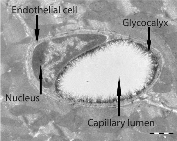
It is possible that the extra-thick glycocalyx barrier lining the capillary lumen of cerebral blood vessels, as shown below, [35] contributes much to a resilient blood brain barrier of low permeability, but then that same glycocalyx may also be vulnerable to the havoc caused by spike protein docking. [36]
The hippocampus is damaged in Alzheimer’s disease
In the case of Alzheimer’s disease, the way that particular damage to the blood brain barrier is thought to unfold is as follows: Brain imaging has shown microbleeds with accumulation of iron residues in the brains of Alzheimer’s patients. The hippocampus is especially vulnerable to and affected by this, in fact far more so than other areas of the brain. [37] The hippocampus is the part of the brain most involved with memory and learning, so it is impacted earlier than other areas, and it is damaged early on in Alzheimer’s disease, with massive die-off of neurons. [38]
Michael Nehls MD PhD writes extensively about injuries to the hippocampus in his book, The Indoctrinated Brain. [39] He traces the injuries to the hippocampus to the discovery by Jonas Salk that adult hippocampal neurogenesis (learning and remembering capacity) is significantly reduced by the cytokine interleukin-6 (IL-6). [40] This is known to happen both by direct neurodegeneration [41] as well as inhibited neurogenesis. [42]
What does this IL-6 have to do with the COVID vaccines? Well, it turns out that spike proteins, such as from coronaviruses, are known to release huge amounts of IL-6 and another highly pro-inflammatory cytokine TNF-α, [43] both of which are potently inflammatory and are thus inhibitors of neurogenesis in the hippocampus, and are therefore antagonistic to an inquisitive, adaptive, learning and remembering brain. So that vulnerability would tend to promote a cognitive impairment or even an Alzheimer’s condition. For that to happen all the more certainly, if a sinister elite wanted to dumb down a population to sheep-like obedience and gullibility, as Dr. Nehls says, “To advance the Great Mental Reset, all that was needed was to ensure that as many people as possible spike regularly – ideally every three to six months – to ensure sustained cerebral toxicity.” [44] He attributes hippocampal damage not only to the repeated bombardment every few months of spike proteins from the COVID vaccines, but also to the following: Lockdowns and social isolation, the carbon-dioxide-laden air inside of masks, lack of exercise from closed gyms and lack of sunlight-provided vitamin D resulting from the most strict lockdowns. “Ultimately, all these factors cause the hippocampus to suffer at any age.” [45]
Cationic lipids deliver the toxic payload, and are also toxic
After COVID injection, the cationic lipids that are used in packaging mRNA are found within the brain in less than 15 minutes, [46] and are also found on autopsy of mRNA vaccine injected deceased individuals. [47] Each of those thousands of packets per dose contain the mRNA instructions to produce spike proteins, and then ACE2 is upregulated.
Those lipids aid the mRNA package to enter cells, and even to cross the blood-brain barrier, due to their outward-facing fat molecules as illustrated here.
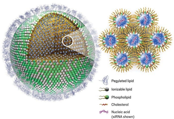
It should also be remembered that the cationic lipids used to envelop mRNA are highly inflammatory and toxic toward the immune system’s macrophages, [49] and those lipids themselves induce pro-inflammatory cytokines TNF-alpha and IL-6. [50] In February 2021, just before the peak vaccine mania heyday, I discussed injuries to liver, lungs and mitochondria that are associated with cationic lipids. This is now the first chapter of my book Neither Safe Nor Effective. Complicating such inflammation, pre-existing inflammatory and auto-immune conditions exacerbate the pro-inflammatory effects of cationic lipids. [51]
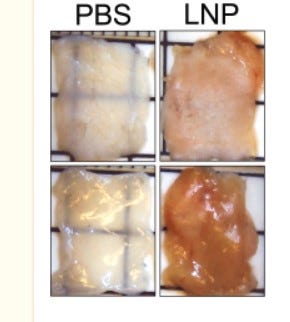
The Ndeupen research team injected mice with empty LNP envelopes, not containing mRNA, and still observed “signs of intense inflammation” with erythema, swelling and “massive and rapid leukocytic infiltrates.” [52] This latter symptom is characteristic of a person having flu-like symptoms or a “bad cold,” and fills the body with suddenly produced white blood cells, as the immune system mounts a desperate response to an invader that is not as easy to attack as the pathogens, such as coronaviruses, that our many generations of ancestors faced. The Ndeupen team found that mortality was significant when higher doses of LNPs were given via intranasal inoculation. If that were known prior to injection of mRNA-carrying LNPs, would any human have consented to injection of these substances?
Placebo (PBS) versus lipid nanoparticle injected (LNP) skin samples from mice show the stark contrast. Note the redness, swelling, opacity and distortion of the LNP-injected skin samples.
How the mRNA vaccines increase risk of cognitive impairment or dementia
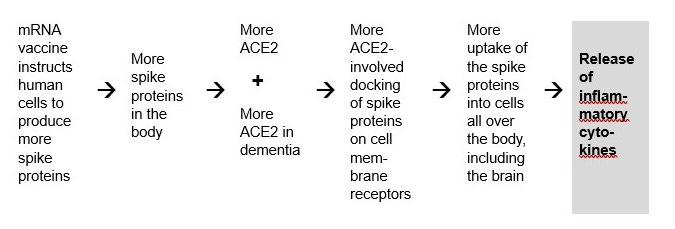
Therefore, the pattern of dementia risk from the COVID vaccines appear to be as follows:
Injection of COVID vaccine dose leads to release of inflammatory cytokines, as follows.
ACE2 plays a key role in inflammation and inflammatory processes, including when spike protein enters by that route. [53] The hyper-inflammatory crisis known as cytokine storm that is seen in severe acute respiratory infections involves abundant release from infected cells of the inflammatory cytokines TNF-α and the interleukins. Furthermore, the higher the levels of these cytokines, the more severe the bout of COVID symptoms and signs. [54]
Neuroinflammation has been correlated with Alzheimer’s disease and other dementia syndromes for nearly a decade. [55]
Spike proteins alone have been shown to interfere with neuronal connectivity and transmission at the synapses. Independently from the Erickson team mentioned above, the Song research team found evidence of neuro-invasion of SARS-CoV-2 into the human brain, and that the ACE2 portal was required for this invasion, and that this was the cause of the neurological symptoms observed. [56]
Thus we see a path from the mRNA COVID vaccines, their spike protein payload and their lipid envelopes, to inflammation as a clinically established risk for, or cause of, dementia and other cognitive impairment.
The Abramczyk research team found evidence of damage to glial cells in the brain following COVID vaccination. [57] Glial cells are supportive cells to neurons, not only giving the physical support of a scaffolding, but electrically as well, maintaining a proper ion balance in the brain. Other glial cells enhance communication among neurons at their synapses and modulate the flow of neurotransmitters.
The way that damage happens to the glial cells following COVID vaccination appears to be as follows: Following Pfizer vaccine dosing, the concentration of cytochrome C decreased in mitochondria. This had the effect of impairing the very essential oxidative phosphorylation pathway in mitochondria, with the resultant unfortunate effect of lower ATP production. The reader may remember that ATP is the unit of currency (so to speak) of energy in the body. It is life-threatening to have any reduction in ATP.
But that was not all of the damage discovered. Lower cytochrome C also results in lower apoptosis, which can lead to cancers, and in fact that malfunction has been noted in one of the deadliest cancers, namely glioblastoma, a particularly difficult brain cancer. Oncologist William Makis MD considers glioblastoma to be one of the most prevalent of the post-COVID vaccine turbo cancers. [58]
If the COVID vaccines have stimulated a mild cognitive impairment among five billion of the earth’s population, how gullible will that population be regarding the next vaccine sales campaign that emerges, and will they be able to remember the casualties from the 2021 COVID vaccine mania heyday?
[1] M Erdogan, O Gurbuz, et al. Prenatal exposure to COVID-19 mRNA vaccine BNT162b2 induces autism-like behavior in male neonatal rats: Insights into WNT and BDNF signaling perturbations. Jan 10 2024. Neurochem Res. 49. 1034-1048. https://link.springer.com/article/10.1007/s11064-023-04089-2#citeas
[2] J Roh, I Jung et al. A potential association between COVID-19 vaccination and development of Alzheimer’s disease. May 28 2024. QJM. https://academic.oup.com/qjmed/advance-article/doi/10.1093/qjmed/hcae103/7684274
[3] Hiroto Komano. Biography. Kakeai. https://kakeai.co.jp/uncategorized/2019/02/06/join_komano/
[4] M Ganaha. Interview of H Komano. Jun 15 2024. https://rumble.com/v51q0r2-mrna.html and on
https://stop-mrna.com/
[5] C Crump, W Sieh. Risk of depression in persons with Alzheimer’s disease: a national cohort study. Apr 14 2024. Alzheimers Dement (Amst). 16 (2). https://www.ncbi.nlm.nih.gov/pmc/articles/PMC11016814/
[6] H Kim, M Kim et al. Psychatric adverse events following COVID-19 vaccination: a populations-based cohort study in Seoul, South Korea. Jun 4 2024. Nature, Molecular Psych. https://www.nature.com/articles/s41380-024-02627-0
[7] I Balasubramanian, A Faheem, et al. Psychiatric adverse reactions to COVID-19 vaccines: A rapid review of published case reports. Apr 13 2022. Asian J Psychiatr. https://www.ncbi.nlm.nih.gov/pmc/articles/PMC9006421/
[8] M Lazareva, L Renemane, et al. New-onset psychosis following COVID-19 vaccination: a systematic review. Apr 11 2024. Frontiers. https://www.frontiersin.org/journals/psychiatry/articles/10.3389/fpsyt.2024.1360338/full
[9] E Rhea, A Logsdon, et al. The S-1 protein of SARS-CoV-2 crosses the blood-brain barrier in mice. Dec 16 2020. Nat Neurosci. 24 (3). https://www.ncbi.nlm.nih.gov/pmc/articles/PMC8793077/
[10] C Huber. The Defeat Of COVID. Apr 2021. https://www.amazon.com/Defeat-COVID-medical-studies-doesnt/dp/0578248212
[11] E Osimo, T Pillinger. Inflammatory markers in depression: A meta-analysis of mean differences amd variability in 5,166 patients and 5,083 controls. Jul 2020. Brain Behav Immun. https://www.ncbi.nlm.nih.gov/pmc/articles/PMC7327519/
[12] M Aminoff, D Greenberg, et al. Clinical Neurology. 6th edition. 2005. Lange McGraw Hill. P. 62.
[13] I Trougakos, E Terpos, et al. Adverse effects of COVID-19 mRNA vaccines: the spike hypothesis. Apr 21 2022. Trends Mol Med. 28 (7). 542-554. https://www.ncbi.nlm.nih.gov/pmc/articles/PMC9021367/#bb0230
[14] A Ogata, C Cheng, et al. Circulating SARS-0CoV-2 vaccine antigen detected in the plasma of mRNA-1273 vaccine recipients. May 20 2021. Clin Infect Dis. https://www.ncbi.nlm.nih.gov/pmc/articles/PMC8241425/
[15] M Erickson, A Logsdon, et al. Blood-brain barrier penetration of non-replicating SARS-CoV-2 and S1 variants of concern induce neuroinflammation which is accentuated in a mouse model of Alzheimer’s disease. Mar 2023. Brain Behav Immun. 109. 251-268. https://www.ncbi.nlm.nih.gov/pmc/articles/PMC9867649/
[16] T Buzhdygan, B DeOre, et al. The SARS CoV-2 spike protein alters barrier function in 2D static and 3D microfluidic in vitro models of the human blood brain barrier. Dec 2020. Neurobiol Dis. 146 (105131). https://www.ncbi.nlm.nih.gov/pmc/articles/PMC7547916/
[17] Z Varga, A Flammer, et al. Endothelial cell infection and endothelitis in COVID-19. Apr 21 2020. Lancet. 395 (10234). https://www.ncbi.nlm.nih.gov/pmc/articles/PMC7172722/
[18] E Kim, M Jeon, et al. Spike proteins of SARS-CoV-2 induce pathological changes in molecular delivery and metabolic function in the brain endothelial cells. Octo 2021. Viruses. 13 (10). https://www.ncbi.nlm.nih.gov/pmc/articles/PMC8538996/
[19] S Raghavan D Kenchappa, et al. SARS-CoV-2 spike protein induces degradation of junctional protein that maintain endothelial barrier integrity. Jun 11 2021. Front Cardiovasc Med. https://www.ncbi.nlm.nih.gov/pmc/articles/PMC8225996/
[20] G Nuovo, C Magro, et al. Endothelial cell damage is the central part of COVID-19 and a mouse model induced by injection of the S1 subunit of the spike protein. Dec 24 2020. Ann Diagn Pathol. https://www.ncbi.nlm.nih.gov/pmc/articles/PMC7758180/
[21] M Frank, K Nguyen, et al. SARS-CoV-2 spike S1 subunit induces neuroinflammatory, microglial and behavioral sickness responses: Evidence of PAMP-like properties. Feb 2022. Brain Behav Immun. 100. 267-277. https://www.ncbi.nlm.nih.gov/pmc/articles/PMC8667429/
[22] X Ou, Y Liu, et al. Characterization of spike glycoprotein of SARS-CoV-2. Mar 27 2020. Nature Commun. 11 (1620). https://www.ncbi.nlm.nih.gov/pmc/articles/PMC7100515/
[23] A Walls, Y Park et al. Structure, function and antigenicity of the SARS-CoV-2 spike glycoprotein. Apr 16 2020. Cell. 181 (2): 281-292. https://www.cell.com/cell/fulltext/S0092-8674(20)30262-2
[24] M Hoffman, H Kleine-Weber, et al. SARS-CoV-2 cell entry depends on ACE2 and TMPRSS2 and is blocked by a clinically proven protease inhibitor. Apr 16 2020. Cell. 181 (2). 271-280. https://www.ncbi.nlm.nih.gov/pmc/articles/PMC7102627/
[25] I Hamming, W Timens, et al. tissue distribution of ACE2 protein, the functional receptor for SARS coronavirus: A first step in understanding SARS pathogenesis. May 7 2004. J Pathol. 203 (2). Pp 631-637. https://www.ncbi.nlm.nih.gov/pmc/articles/PMC7167720/
[26] T Buzhdygan, B DeOre, et al. The SARS CoV-2 spike protein alters barrier function in 2D static and 3D microfluidic in vitro models of the human blood brain barrier. Dec 2020. Neurobiol Dis. 146 (105131). https://www.ncbi.nlm.nih.gov/pmc/articles/PMC7547916/
[27] G Nuovo, C Magro, et al. Endothelial cell damage is the central part of COVID-19 and a mouse model induced by injection of the S1 subunit of the spike protein. Dec 24 2020. Ann Diagn Pathol. https://www.ncbi.nlm.nih.gov/pmc/articles/PMC7758180/
[28] Ibid Buzhdygan et al.
[29] B DeOre, K Tran, et al. SARS-CoV-2 spike protein disrupts blood-brain barrier integrity via RhoA activation. Oct 21 2023. J Neuroimmune Pharmacol. 16 (4). 722-728. https://www.ncbi.nlm.nih.gov/pmc/articles/PMC8536479/
[30] E Rhea, A Logsdon, et al. The S1 protein of SARS-CoV-2 crosses the blood brain barrier in mice. Dec 16 2020. Nat Neurosci. 24 (3). 368-378. https://www.ncbi.nlm.nih.gov/pmc/articles/PMC8793077/
[31] Ibid Buzhdygan et al.
[32] M Erickson, A Logsdon, et al. Blood-brain barrier penetration of non-replicating SARS-CoV-2 and S1 variants of concern induce neuroinflammation which is accentuated in a mouse model of Alzheimer’s disease. Mar 2023. Brain Behav Immun. 109. 251-268. https://www.ncbi.nlm.nih.gov/pmc/articles/PMC9867649/
[33] A Montagne, S Barnes, et al. Blood-brain barrier breakdown in the aging human hippocampus. Jan 21 2015. 85 (2). 296-302. https://www.ncbi.nlm.nih.gov/pmc/articles/PMC4350773/
[34] C Iadecola. Neurovascular regulation in the normal brain and in Alzheimer’s disease. May 1 2004. 5. 347-360. Nature Rev Neurosci. https://www.nature.com/articles/nrn1387
[35] R Haeren, S van de Ven, et al. Assessment and imaging of the cerebrovascular glycocalyx. Apr 14 2016. Current Neurovasc Res. https://www.researchgate.net/publication/301896448_Assessment_and_Imaging_of_the_Cerebrovascular_Glycocalyx/download
[36] P Stoddart, S Satchell, et al. Cerebral microvascular endothelial glycocalyx damage, its implications on the blood-brain barrier and a possible contributor to cognitive impairment. Apr 1 2022. Brain Res. https://www.sciencedirect.com/science/article/abs/pii/S0006899322000282
[37] A Montagne. Ibid. https://www.ncbi.nlm.nih.gov/pmc/articles/PMC4350773/
[38] Y Mu, F Gage. Adult hippocampal neurogenesis and its role in Alzheimer’s disease. Dec 22 2011. Mol Neurodegen. 6 (85). https://www.ncbi.nlm.nih.gov/pmc/articles/PMC3261815/
[39] M Nehls. The Indoctrinated Brain. 2023. Skyhorse Publishing. Pp. 88-89.
[40] L Vallières, I Campbell, et al. Reduced hippocampal neurogenesis in adult transgenic mice with chronic astrocytic production of interleukin-6. Jan 15 2002. https://www.ncbi.nlm.nih.gov/pmc/articles/PMC6758670/
[41] A Samuelsson, E Jennische, et al. Prenatal exposure to interleukin-6 results in inflammatory neurodegeneration in hippocampus with NMDA / GABA-a dysregulation and impaired spatial learning. May 1 2006. Dev Psych Pregnancy. https://journals.physiology.org/doi/full/10.1152/ajpregu.00268.2005
[42] A Mouihate, S Kalakh. Maternal interleukin-6 hampers hippocampal neurogenesis adult rat offspring in a sex-dependent manner. May 21 2021. Dev Neurosci. https://karger.com/dne/article-abstract/43/2/106/107656/Maternal-Interleukin-6-Hampers-Hippocampal
[43] W Wang, L Ye, et al. Up-regulation of IL-6 and TNFα induced by SARS coronavirus spike protein in murine macrophages via NF-ƙB pathway. Sep 2007. 128 (1). 1-8. https://www.ncbi.nlm.nih.gov/pmc/articles/PMC7114322/
[44] M Nehls, The Indoctrinated Brain. Ibid. p. 108.
[45] Nehls, The Indoctrinated Brain. Ibid. p. 116
[46] SARS-CoV-2 mRNA vaccine. Pfizer report, Japanese government. [Document in both Japanese and English]. P. 7 of the English section. https://www.docdroid.net/xq0Z8B0/pfizer-report-japanese-government-pdf
[47] N Hulscher, P Alexander, et al. A systematic review of autopsy findings in deaths after COVID-19 vaccination. Pre-proof. Jun 17 2024. https://doi.org/10.1016/j.forsciint.2024.112115
[48] R Cross. Without these lipid shells, there would be no mRNA vaccines for COVID-19. Mar 6 2021. Chem and Eng News. https://cen.acs.org/pharmaceuticals/drug-delivery/Without-lipid-shells-mRNA-vaccines/99/i8
[49] M Filion, N Phillips. Toxicity and immunomodulatory activity of liposomal vectors formulated with cationic lipids toward immune effector cells. Oct 23 1997. Biochimica et Biophysica Acta. 1329 (2). https://www.sciencedirect.com/science/article/pii/S0005273697001260
[50] Y Zhu, Y Meng, et al. Toxicological exploration of peptide-based cationic liposomes in siRNA delivery. Jul 1 2019. Colloids Surface B Interfaces. Vol 179 66-76. https://www.sciencedirect.com/science/article/abs/pii/S0927776519302012?via%3Dihub
[51] I Trougakos, E Terpos, et al. Adverse effects of COVID-19 mRNA vaccines: the spike hypothesis. Apr 21 2022. Trends Mol Med. 28 (7). 542-554. https://www.ncbi.nlm.nih.gov/pmc/articles/PMC9021367/#bb0230
[52] S Ndeupen, Z Qin, et al. The mRNA-LNP platform’s lipid nanoparticle component used in preclinical vaccine studies is highly inflammatory. Nov 20 2021. 24 (12). iScience. https://www.ncbi.nlm.nih.gov/pmc/articles/PMC8604799/
[53] M Iwasaki, J Saito, et al. Inflammation triggered by SARS-CoV-2 and ACE2 augment drives multiple organ failure of severe COVID-19: Molecular mechanisms and implications. Oct 8 2020. Inflammation. https://www.ncbi.nlm.nih.gov/pmc/articles/PMC7541099/
[54] Q Ruan, K Yang, et al. Clinical predictors of mortality due to COVID-19 based on an analysis of data of 150 patients from Wuhan, China. Mar 3 2020. Intensive Care Med. https://www.ncbi.nlm.nih.gov/pmc/articles/PMC7080116/
[55] V Calsoloaro, P Edison. Neuroinflammation in Alzheimer’s disease: Current evidence and future directions. May 11 2016. Alzheimer’s & Dementia. https://alz-journals.onlinelibrary.wiley.com/doi/full/10.1016/j.jalz.2016.02.010
[56] E Song, C Zhang, et al. Neuroinvasion of SARS-CoV-2 in human and mouse brain. Jan 12 2021. J Experim Med. 218 (3). https://rupress.org/jem/article/218/3/e20202135/211674/Neuroinvasion-of-SARS-CoV-2-in-human-and-mouse
[57] H Abramczyk, B Brozek-Pluska, et al. Decoding COVID-19 mRNA vaccine immunometabolism in central nervous system: human brain normal glial and glioma cells by Raman imaging. Mar 2 2022. bioRxiv. https://www.biorxiv.org/content/10.1101/2022.03.02.482639v1.full
[58] W Makis. mRNA injury series – Turbo brain cancer due to university COVID-19 vaccine mandates in the United States – three tragic cases. Feb 15 2024. MakisMD.


